Lifelike tissues that predict human outcomes
Successful translation demands biology that behaves exactly like a patient's own. We grow human organ tissues so intricate they recreate phenotype down to the cell.


Interventional studies at wafer-scale
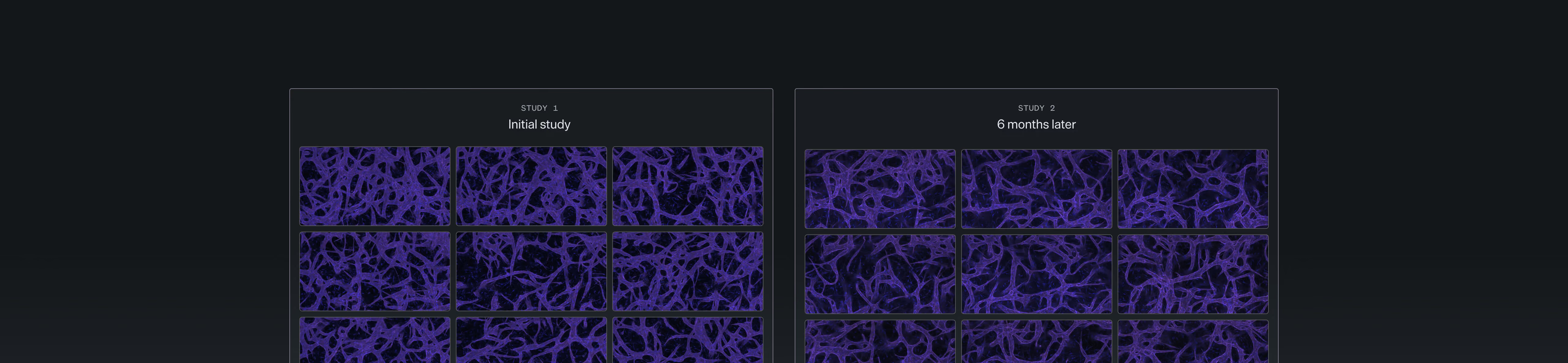
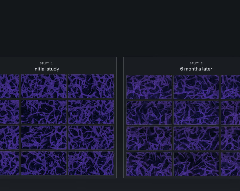
Fully perfusable vascular networks
A complete tissue microenvironment
Reproducible batch after batch
Each of our tissues contains 200,000 to 500,000 individual cells, faithfully reconstituting the phenotypes and functions of native human tissues. Diverse donor sourcing lets us capture the biological variation seen across patient populations.
Tissue-specific, self-assembled 3D vascular networks can be fully perfused, enabling intravascular administration of therapeutics with realistic physiological transport.
Epithelium, endothelium, and stroma organize with both innate and adaptive immunity, wired together by the same biochemical and mechanical signals found in the body.
Grown in Vivodyne-optimized formulations and handled end-to-end by our HIVE robotic platform, our tissues form with stunning consistency across experiments.
20+ human tissue models Hundreds of clinical applications
Our suite of human tissue models span the most critical areas in medicine. Each tissue model is available in healthy and diseased states.
Our bone marrow-on-a-chip facilitates the production of blood cells needed to oxygenate tissues, regulate hemostasis, and provide immunity.
The multi-lineage development of myeloid and erythroid cells from hematopoietic stem and progenitor stem cells (HSPCs) recapitulates the native process of hematopoiesis.
This model supports the screening of anti-cancer drugs for hemotoxicity and the modeling of disease states such as erythropenia, anemia, neutropenia, and immunosuppression.

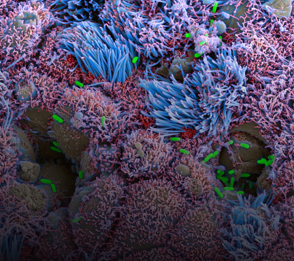
Our large and small airway models replicate the hallmark features of the bronchi and bronchioles within the lung, comprising beating ciliated cells, mucus producing goblet cells, mature epithelial basal cells and protective club cells.
Lung cell differentiation and mucociliary beating is achieved via culture with an air-liquid interface.
The model faithfully recapitulates neutrophil extravasation in response to inflammatory stimuli such as bacterial infection, cigarette smoke, aerosolized particles, and viruses, and can be leveraged to model chronic obstructive pulmonary disease (COPD).
Our liver-on-a-chip enables deep analysis of native liver functions such as drug metabolism, protein synthesis, waste clearance and blood glucose regulation.
The fully vascularized liver model supports urea synthesis and albumin production, the formation of robust bile canaliculi, quantification of drug-induced liver toxicity (DILI) biomarkers such as alanine aminotransferase (ALT), and high-content secretome analysis.
This is a ready-to-deploy platform for predicting DILI and drug metabolism and modeling diseases such as hepatitis, fatty liver disease and cirrhosis.

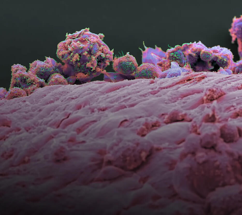
In our solid-tumor models, co-cultures of tumor spheroids with parenchymal and stromal cells provide a physiologically relevant niche for modeling cancer biology.
By incorporating key components of the tumor microenvironment, we model the microarchitecture of cancerous tissue, tumor growth, angiogenesis, epithelial-mesenchymal transition (EMT), and metastasis.
The solid-tumor-on-a-chip enables screening of anticancer drugs and next generation immunotherapies.
Our islet-on-a-chip captures the pancreas’ ability to secrete enzymes for digestion and produce hormones for regulating blood glucose levels. The model recapitulates in vivo glucose-stimulated insulin secretion and incorporates all the major hormone producing endocrine cells (alpha, beta, delta, and PP cells) of the pancreas.
This platform supports testing and screening of diabetes drugs for enhancing insulin release and may be leveraged to monitor autoimmune pathogenesis and beta cell destruction.
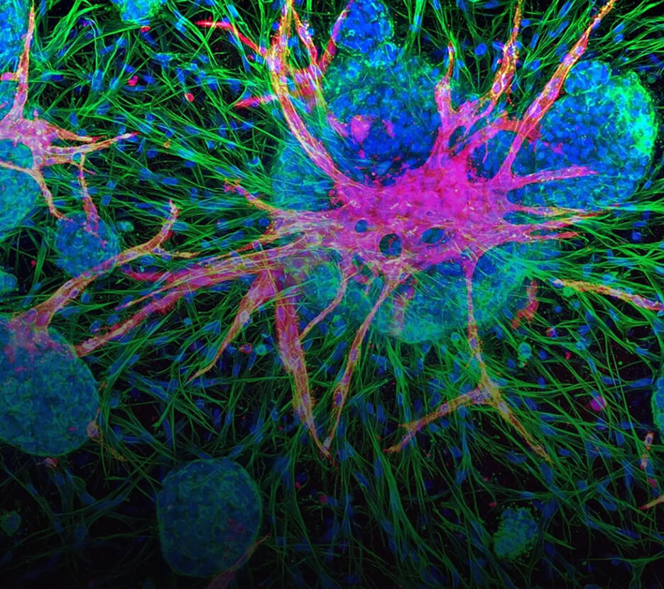
Our intestine-on-a-chip encompasses villi-like structures and the major intestinal cell types including enterocytes, enteroendocrine cells, goblet cells, and paneth cells.
The formation of mature enteroids is achieved via epithelial barrier formation with intestinal barrier function, mucus production, and the development of complex gut-microbiota, all of which contribute to the intestine’s role in food digestion and nutrient absorption.
The model reproduces key functions of the intestine, pathological features of IBD and innate immune responses, allowing for the testing of drug pharmacokinetics/pharmacodynamics (PK/PD), modeling of inflammatory and infectious diseases, and screening of drug compounds for gastrointestinal toxicity.

Our model mimics the bi-layered architecture and hemodynamic environment of the native placenta barrier. This regulates the transport of materials between the mother and the fetus during pregnancy.
The model incorporates cytotrophoblasts, which undergo syncytialization to form a cell monolayer with tight intercellular junctions, fetal endothelial cells separated by a basement membrane, and microvilli on the apical surface, which serve to increase the surface area of the maternal-fetal interface and act as sites for the expression of active membrane transporters associated with drug and xenobiotic transfer.
Leverage this to evaluate the PK/PD of maternally administered drugs to improve preclinical evaluation of drug safety in pregnancy.
This model recapitulates the adhesion of embryos to the endometrium and subsequent invasion into the maternal tissue, mimicking early implantation and placentation observed in early pregnancy.
We reconstruct the three-dimensional structural organization of the maternal-fetal interface, simulating the invasion of trophoblasts into the maternal uterus and the subsequent arterial remodeling that is necessary for improved blood circulation.
This platform provides insights into implantation abnormalities and disorders of placentation, enabling the development of therapeutics for early pregnancy and the evaluation of drug compounds' off-target effects.
.jpg)
Our skeletal muscle-on-a-chip recapitulates the anisotropic structure and mechanical properties of native muscle, comprising of realistic muscle that undergoes stimulated contraction.
We establish a microenvironment that supports the myogenic differentiation of mesenchymal stromal cells and the formation of aligned multinucleated myotubes and myofibers with directional organization.
This platform faithfully models oxidative muscle injury and cachexia, enabling screening of therapies for muscle injuries or drug toxicity against skeletal muscle.

The blood-brain barrier (BBB)-on-a-chip comprises specialized microvascular networks, mural cells, and astrocytes that together mimic the barrier function of the human BBB to separate blood from brain interstitial fluids.
Small molecule and immune cell transport across the BBB model is regulated by tight junctions, endothelial cell-expressed transporters, transcytosis, leukocyte adhesion molecules, and signaling across the incorporated cell populations.
This platform can be leveraged to screen the transport of drugs targeted to the brain for neurological and psychiatric diseases and to model BBB dysfunction implicated in diseases such as multiple sclerosis, stroke and epilepsy.

Human data, before the clinic
We work collaboratively with leading biopharmas to advance our shared mission to bring breakthrough medicines to patients in need.

